|
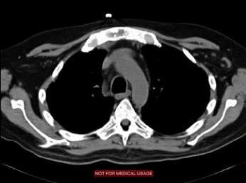
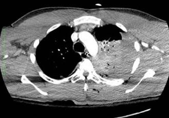
The patient on the right has MUCH more muscle mass, and very little fat, compared to our patient. He looks like a body-builder. He was given IV contrast, as his aorta and superior vena cava are much higher in attenuation than muscle. This is again an axial or horizontal image, displayed with soft tissue windows. He had trauma and his left lung and left pleural space are partly full of blood. Can you tell on our patient (on the left) whether he has pneumonia? Recall that he presented with cough and fever.
|
|