ICM-II: CHEST, case 4 |
||
 |
|||||||||||
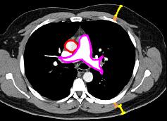
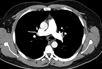
This is a chest CT scan, axial plane, with IV contrast, displayed with soft tissue window. The contrast is brighter in the pulmonary artery than the aorta, so the scan was begun early after contrast injection, to maximize contrast in the pulmonary portion of the vasculature. This is a good way to identify centrally located pulmonary emboli. This is a normal scan, not from our patient. There is a bit more fat than muscle in the pectoral and back regions, suggesting mild obesity.
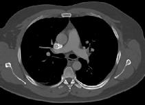
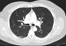
What are these two other images? Did the patient receive additional radiation for these images?