|
SUMMARY
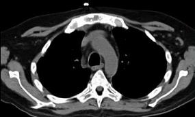
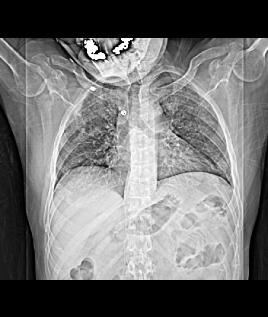
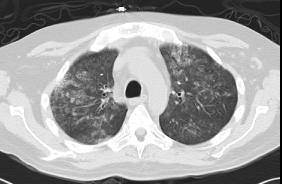
To summarize, we reviewed again how to estimate body habitus on a frontal view of the chest, the concept of a CT topogram, as well as some technical factors to check on cross-sectional imaging, such as how to identify a CT, the imaging plane, the window, and whether contrast was used. Recall that on a CT, the cortical part of bones are the whitest thing on the image, and that on a soft tissue window, fat and muscle will be very different shades of grey. For detection of IV contrast, look for a big vessel like the aorta and compare its attenuation to muscle. If it is whiter than muscle, then IV contrast is present.
|
|