Tufts Anatomy Yr-1 |
||||||||||
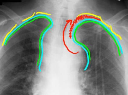
If you look closely at the two lung apices, the left one looks lower than the right. This is another sign of significant bleeding from central vessels. The blood can dissect out from the mediastinum (red arrow) and peel the parietal pleura (blue) off the inside of the ribs (yellow), collecting in a crescent-shaped rim and displacing lung and visceral pleura (green) downward. This appearance is called an apical ‘cap’. The blood is NOT in the pleural space, but in the extrapleural space, a potential connective tissue space between the ribs and the parietal pleura.
A
B
CASE 1 followup