Tufts Anatomy Yr-1 |
||||||||||
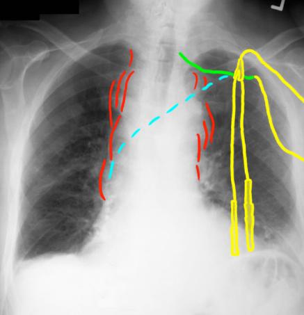
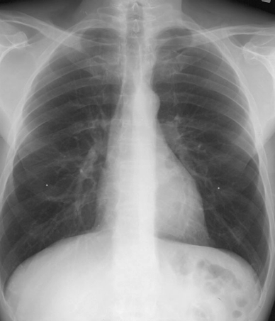
The mediastinum in this patient (Image A) is markedly abnormal, wide and irregular, as shown here in red. The central line is NOT traveling toward the superior vena cava as expected (normal course is shown in blue). The portion of the line that is outside the patient is again shown in yellow, and the portion that is inside the patient is shown in green. A normal chest radiograph (CXR) is shown in Image B.
A
B
CASE 1 followup