Anatomy |
||||||||||||||||||||
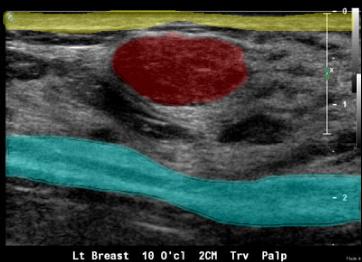
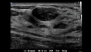
This breast US shows a solid mass that is wider than tall with smooth rounded margins. The skin is shown in yellow and the pectoral muscles in blue. The image field is a rectangle, consistent with use of a linear high frequency small parts transducer. This abnormality is most likely a fibroadenoma, which could either be followed (in a young patient) or biopsied with ultrasound guidance.
A