ICM-II: ABDOMEN, case 4 |
||
 |
||
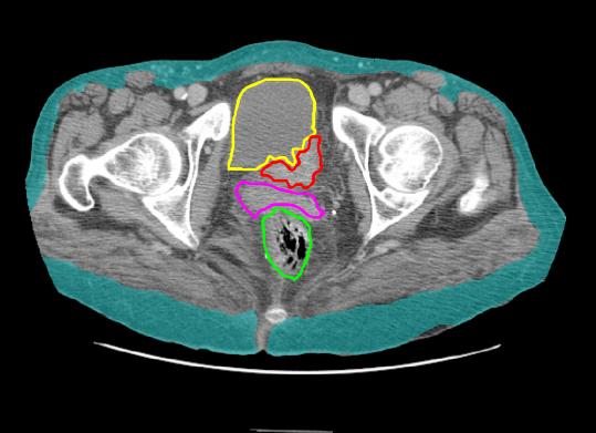
This is a CT image (bone is very white), and the patient has a relatively normal amount of subcutaneous fat. The rectum and vagina are normal. The bladder is indented on the left near the region of the uretero-vesical junction (UVJ) by a tumor. This is causing the patient's symptoms and the ultrasound finding of left hydronephrosis. What other type of imaging might be used to evaluate this tumor? |
||
 |
|||||||||||