Anatomy Yr-1 |
||||||||||||||
65 year old female, left shoulder pain |
||||||||||
 |
||||
 |
||||
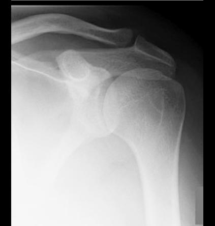
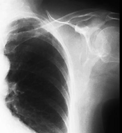
The lungs in our patient are much smaller than the normal comparison, suggesting that she has not taken a deep breath. Image A is a closeup of our patient's left shoulder from the chest radiograph and Image B is a normal left shoulder radiograph. Positioning of the shoulder bones on a chest radiograph is usually not optimal. Do you seen anything abnormal, focusing on the glenoid? |
||||||
B
A