Anatomy Yr-1 |
||||||||||||||
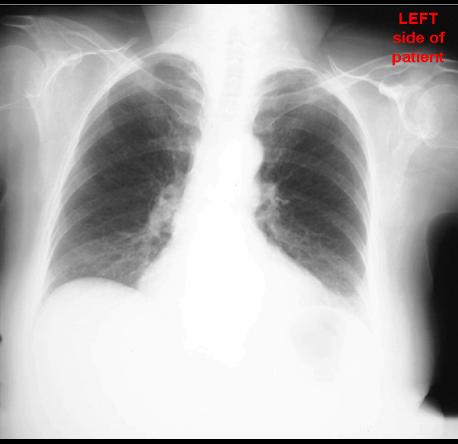
65 year old female, left shoulder pain |
||||||
 |
 |
|||||
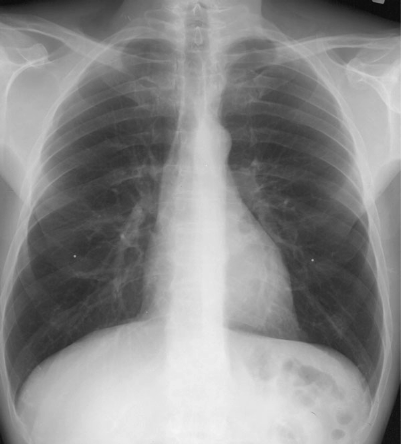
Image A is our patient and Image B is a normal comparison on a different patient. For radiographs, regardless of how the beam passed through the patient (from front --> back or from back --> front), we always display the image as if we are FACING the patient. So the LEFT side of the patient's body is shown on the RIGHT side of the image. Given this normal comparison, what do you think of the patient's lungs? Do we have a good view of their left shoulder? |
||||||
B
A