Tufts Anatomy Yr-1 |
|||||||||||||||||
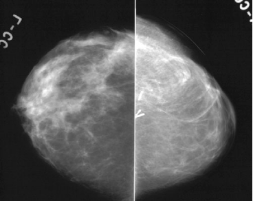
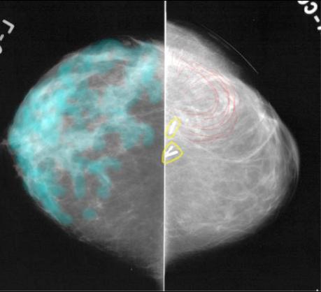
43 year old woman with right breast cancer and prior lumpectomy with reconstruction. |
||||||||||
 |
||||||||||
Just like in other parts of the body, when imaging the breast we like to get images from at least two different angles. The views shown in Image A are called 'CC' or cranio-caudal views. They are top-down views. |
||||||||||
 |
||||||||||
When looking at mammograms we focus on symmetry between the two sides. As on the previous MLO views, the breasts do not look symmetric on the CC views. In Image B, the normal tissue of the breast is shown in aqua on the left. In the right breast, we can see a few surgical clips again (circled in yellow) and that unusual swirling linear pattern, shown in red. Can you think of a tissue of the body that could have been used for the patient's breast reconstruction that might have this appearance on x-ray? |
||||||||||
B
A