Tufts Anatomy Yr-1 |
||||||||||||||||||||
At the end of each case is a 'followup' link to additional related images and cases. These are for you optional review, and are not required prior to the class session. |
||||||||||||||||||||
 |
||||||||||||||
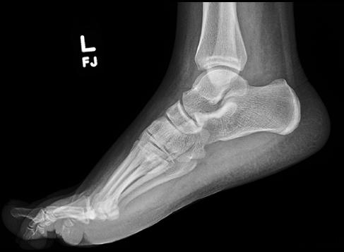
This is the normal lateral view, so that you can practice locating the bones of the foot. Try to identify them before using the labels. |
||||||||||||||
Other types of imaging that could be done to look for signs of bony infection include MR, CT or bone scanning. To review the most approriate types of imaging that can be done in this setting, click the link below to review the American College of Radiology Appropriateness Criteria (a free online resource, organized by clinical symptoms). |
||||||||||||||