PrISM Cardiovascular |
||||||||||||||||||||||||
 |
||||||||||||||||||||||||
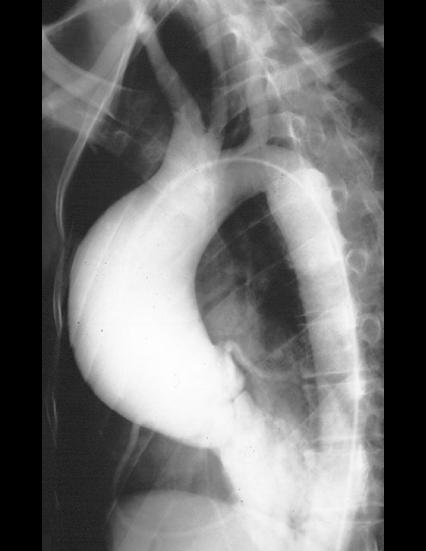
There are many different ways to evaluate the aorta and aortic valve with imaging, including CT, MR, and echo. This patient had angiography, which would NOT be the initial study if the case were done today, since it is an invasive study with a high radiation and contrast dose, but it does show the anatomy well. Try to find the listed structures on your own before mousing over the labels. |
||||||||||||||||||||||||
This patient was found to have Marfan's syndrome and a dilated aorta because of this condition, with regurgitation through the aortic valve, since the valve cusps could no longer close tightly as the diameter of the aorta increased.