PrISM Cardiovascular |
|||||||||||||||||||
 |
|||||||||||||||||||
What other imaging could be done in this patient to figure out what is going on? |
||||||
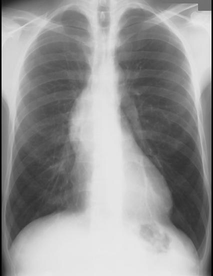
There is a bulge along the right mediastinal border that looks unusual. One possible cause for such a bulge would be enlargement of the ascending aorta. The rest of the aorta does not look enlarged, so this is unlikely to be a diffuse aneurysm.
When only the proximal aorta is enlarged, there are several possible causes, which could include certain infections (syphilis), but also connective tissue disorders that may weaken the wall, as in Marfan's syndrome. This patient does look VERY tall, so this is a possibility.
Another possibility is aortic stenosis, where the force of blood coming through the narrowed valve can hit the ascending aortic wall, causing it to develop a bulge in just this place, as shown on the 'force' label. This is called 'post-stenotic dilatation'.
Obviously, physical exam findings would be very helpful in this case, including examination for elongated fingers, and auscultation of the heart for murmurs.