PA Anatomy: Chest: Case 3 |
||||
 |
|||||||||||||
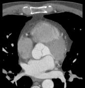
The study on the previous page was a chest CT scan, displayed with a soft tissue window. There is intravenous contrast present, mostly in the aorta and left atrium, so imaging was slightly late after injection. A single image from the series is shown below. Note that the blood vessels are whiter than body wall muscles, and cortical bone is the whitest area on the image. |
|||||||||||||
 |
|||||||||||||
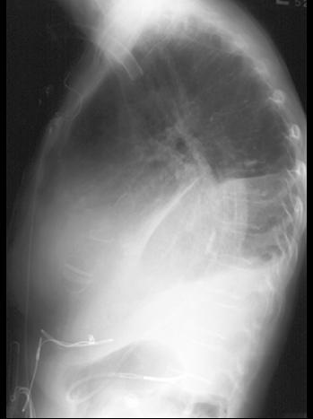
Using your knowledge of orientation of cardiac chambers shown on CT, mouse over the various chambers to locate them on the lateral CXR above. Can you decide which valves were replaced? |
|||||||||||||