Anatomy |
||||||||||||||||||||
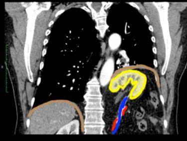
This patient has a congenital diaphragmatic hernia, called a Bochdalek hernia. In this case, it is a very large hernia, with most of the left kidney displaced upward into the chest, stretching the renal artery and vein. The diaphragm on the left is displaced upward. Image B is a select image from Patient A, and Image C shows colored outlines on key structures. Because the embryology of the diaphragm is complex, weak spots may result from incomplete fusion of precursor tissues. If the weakness is posterior in location, a Bochdalek hernia can develop. If the weakness is anterior, a different hernia called a Morgagni hernia can result.