Anatomy |
||||||||||||||||||||
 |
||||
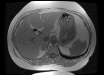
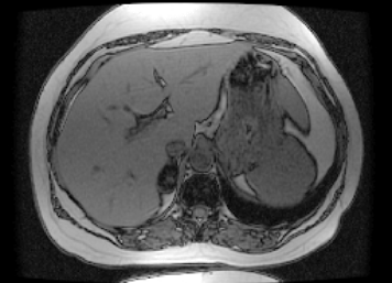
Image A is a T1-weighted MR image (note the dark CSF, outlined in aqua), aorta, IVC and right adrenal mass. Image B is an out-of-phase image, a special MR sequence that is sensitive to fat within masses. You can recognize this sequence from the 'India ink' artifact that makes all structures look like they have a blackoutline around them. |
||||
 |
||||
A
B
The right adrenal mass has become quite dark on Image B, indicating that it contains fat, and making a benign adenoma the most likely diagnosis. Overall adenomas are a more common cause of unilateral adrenal masses than metastases.