Anatomy |
||||||||||||||||||||
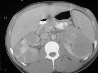
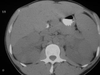
Image A is a CT without IV contrast (but with GI contrast), and Image B is with IV contrast.
On Image A, you can tell that the kidneys look large and lumpy but not much else.
On Image B, you can see that there are many masses in both kidneys. This is not the typical presentation for renal cell carcinoma, which is most often unilateral.
In this case, the patient has renal lymphoma, also rare.
Patients with genetic syndromes like von Hippel Lindau or tuberous sclerosis can develop multiple bilateral renal tumors that might look similar to this case.
A
B
Renal abscesses might also be multiple and bilateral. If you are worried about a renal infection, it can sometimes be helpful to look at CT images on a LUNG window to try to detect tiny amounts of gas.