Anatomy |
||||||||||||||||||||
 |
||
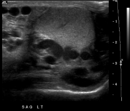
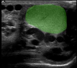
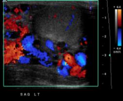
This is a scrotal ultrasound (the testicle is shown in green in Image B), with many tortuous anechoic round and tubular structures consistent with dilated veins. There is a lot of flow on the color Doppler image (Image C), consistent with a varicocele. Thinking of the anatomy of the region, how could a flank mass cause this? |
||
A
B
C