Anatomy |
||||||||||||||||||||
 |
||
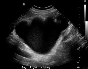
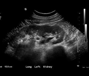
The right kidney on Image A is very abnormal, with just an extremely dilated collecting system and no renal parenchyma visible. The left kidney on Image B is normal in appearance, with a thick cortex and echogenic renal sinus containing vessels and possibly some normal parts of the collecting system. The upper pole is always to the left on sagittal images. This is an example of post-obstructive atrophy on the right--the kidney parenchyma thins and is destroyed when high-grade obstruction is not relieved promptly.
A
B
head
head
feet
feet