Anatomy |
||||||||||||||||||||
 |
||||
 |
||||
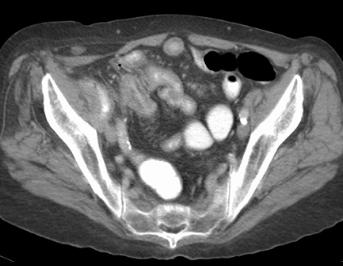
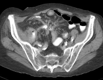
There is an abscess (in pink on the labeled psoas images) in the right psoas muscle, containing contrast and gas (yellow arrow on labeled psoas images). Findings like stranding and distortion of the mesentery as in this case, and thickening of the small bowel wall would not be visible on a KUB or fluoroscopic contrast study. For such reasons, CT is preferred for many clinical situations in which fluroscopic contrast studies were previously done.