Anatomy |
||||||||||||||||||||
 |
||
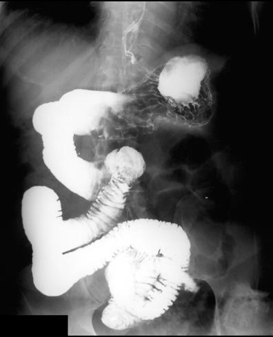
This study is a small bowel follow-through, which has been replaced in most clinical settings with CT, either with positive contrast (barium or water-soluable contrast) or negative contrast (water or other radio-lucent liquid).
No patient preparation is needed for this study. The patient drinks barium and fluoroscopy with sequential images are obtained as the contrast enters different loops of small bowel.
In this patient, the proximal small bowel loops are all on the right side of the abdomen, and the ligament of Treitz is not in the normal position, suggesting that the patient has malrotation, a congential abnormality unrelated to inflammatory bowel disease.
The study does show the normal ruffled appearance of the rugae in the stomach, and the normal, closely positioned, evenly-spaced plicae of several proximal loops of small bowel with smooth outer margins.
The blue button links to ACR recommendations for evaluation of Crohn disease.