Anatomy |
||||||||||||||||||||
 |
||
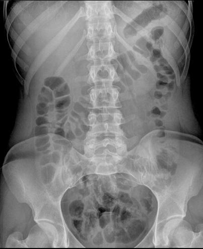
This is a normal KUB in another patient--try to use the features from the prior table to determine the location of the stomach, small intestinal loops and colon before showing the labels.
Because they are normally filled with mostly fluid, small intestinal loops are often hard to find on a KUB and their folds are often not very apparent.
The portion of the colon that is often the easiest to find on a supine KUB is the transverse colon. Why would this part of the colon be particularly likely to be filled with gas on such an image?
As in the previous table, the labels on this image show the stomach in yellow, the small intestine in blue and the colon in pink.