Anatomy |
||||||||||||||||||||
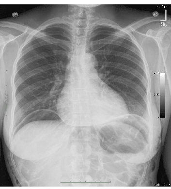
This image shows free air in the abdomen. The radiograph is a chest x-ray, not a KUB. The centering of the diaphragms on the image is better on a CXR than a KUB when the question is possible perforation. This was done in the upright position, so the air has risen to be sandwiched between the bottom of the diaphragm and the top of the liver on the right, spleen on the left. This gives the most sensitive view for detecting small amounts of free air. This is a LOT of free air. The next radiographic study depends entirely on the patient's symptoms and history. If she is acutely ill, NO further imaging is indicated and she should go straight to the OR! |
|||||||||
 |
|||||||||