Anatomy |
||||||||||||||||||||
 |
||||
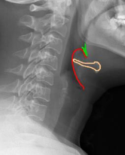
This is a lateral radiograph of the neck, which is sometimes done prior to other more definitive studies to look at the airway. |
||||
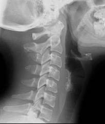
In Image A from our patient, the hyoid bone is shown in orange, and a very thick and inflamed epiglottis in red. The region of the valleculae is shown in green. This is an emergency, as epiglottitis can lead to complete airway obstruction. Image B is a comparison study to show the normal appearance of the epiglottis. |
||
A
B