Anatomy |
||||||||||||||||||||
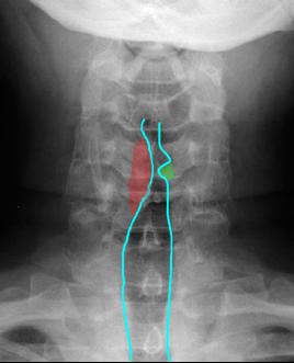
This is an AP radiograph of the neck, showing asymmetry in the air in the region of the larynx. The image was obtained during phonation, so the normal left vocal cord is adducted (green), while the contour on the right is abnormal (red), suggesting a mass and a reason for hoarseness.
This is a CT at the level of the vocal cords. Do you think it is normal? An understanding of anatomy is vital when looking at neck CT. Try to identify the vascular structures and figure out what laryngeal structures we are seeing.