|
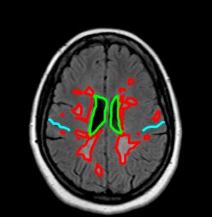
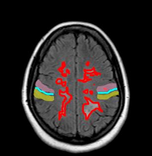
What type of study is this? This is a head MRI, axial plane. The pulse sequence is a special type of sequence called FLAIR, which is a type of T2 weighting that shows water in the CSF as low signal, but other areas of abnormal water in tissues as HIGH signal (red on the labeled images). This is a great way to see abnormalities that are immediately adjacent to the ventricles or sulci, which on a regular T2 sequence might be hard to separate from the high signal from the CSF. How would you describe the abnormal findings? There are areas of abnormal high signal located in the white matter adjacent to the ventricles and extending outward and superiorly on both sides. These locations would be described as periventricular, deep and subcortical white matter. What is labeled in aqua, pink and yellow on these images?
|
|