Imaging Pneumonia |
Case 9-Answers |
|||||||||||||
 |
||||||||||||||
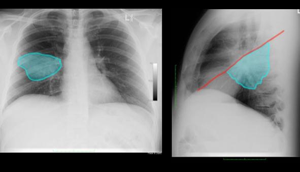
This opacity is in the middle of the right lung on the frontal view, suggesting that it is either in the RML or RLL. The lateral view shows it to be posterior in location, so in the right lower lobe. |
||||||||||||||
The lateral view is key to figuring this case out, since the frontal does not localize the lesion completely. It does not abut either the heart or diaphragm, so you cannot use these features on the frontal view to help decide where it is. On the lateral, it is posterior to the major fissure (red), so in the uppermost part of the RLL. |
||||
 |
||||