Imaging Pneumonia |
|||||||||||||
Case 8-Answers |
|||||||||||||
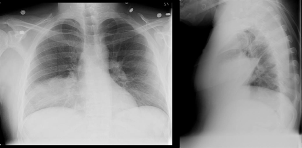 |
This large opacity is in the lower right chest on the frontal view, and anterior, overlying the heart on the lateral view, consistent with another right middle lobe pneumonia. |
||||||||||||
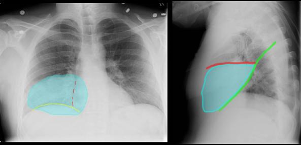
The opacity fills the entire right middle lobe region on the lateral view, between the minor fissure (red) and the major fissure (green). |
||||||
The opacity completely obscures the right heart border (red dotted line) but not the right hemidiaphragm (yellow). |
||||||
 |
||||||