Imaging Pneumonia |
||||||||||||||
Case 6-Answers |
||||||||||||||
 |
||||||||||||||
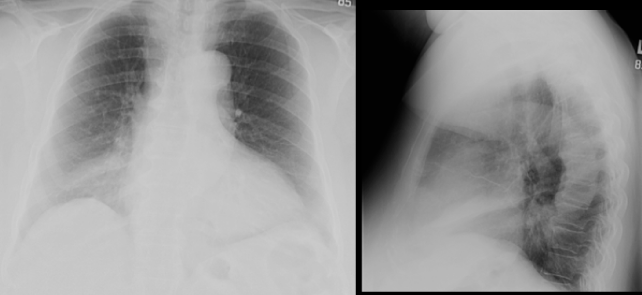
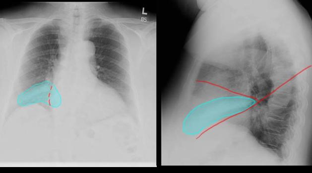
This opacity is a little smaller and harder to see than some of the other cases, but is shown in the lower right chest on the frontal view, and overlying the heart on the lateral view, in the region of the right middle lobe. |
||||||||||||||
 |
||||||
The consolidation obscures a part of the right heart border slightly (red dotted line-compare sharpness of right to left border) but does not obscure the right hemidiaphragm. |
||||||
On the lateral view, the opacity is seen between the minor and major fissures, shown in red. |
||||||