Imaging Pneumonia |
Case 5-Answers |
|||||||||||||||
 |
||||||||||||||||
 |
||||||||||||||||
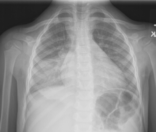
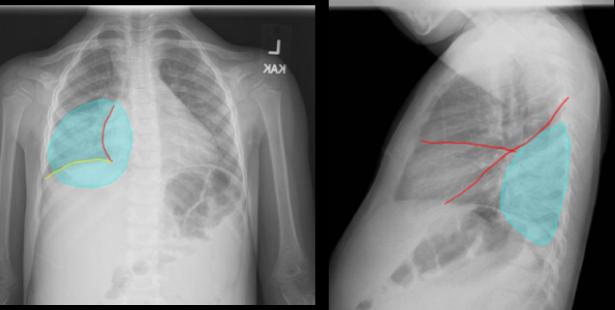
This is another RLL pneumonia that is slightly higher in position than some of the other ones we have seen. It is in the lower posterior right part of the chest. |
||||||||||||||||
 |
||||||
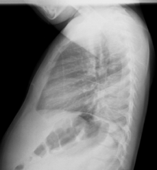
On the lateral view, it is obscuring just the most posterior bit of the hemidiaphragm, and is positioned posterior to the major and minor fissures, shown in red. |
||||||
Because this area of consolidation is slightly high within the RLL, it is not obscuring either the heart border (red) or the right hemidiaphragm on the frontal view (yellow). |
||||||