Imaging Pneumonia |
Case 2-Answers |
|||||||||||||||
 |
||||||||||||||||
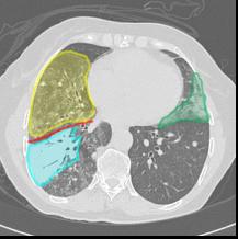
There is opacity posteriorly and inferiorly on the right most consistent with RLL pneumonia. On CT, the densest area of consolidation is in the right lower lobe. |
||||||||||||||||
 |
||||||||||||
 |
||||||||||||
 |
||||||||||||
CT is more sensitive than radiography for finding areas of subtle pneumonia, and there is increased density in other lobes on the CT, including the right middle lobe (yellow) and the lingula (green). The major fissure is shown in red. The most dense consolidation in the right lower lobe is shown in blue. |
||||||||||||
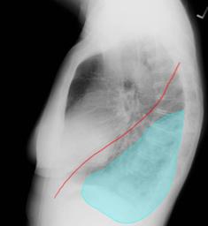
The opacity is posterior in location, making the spine appear denser than normal. The major fissure is shown in red.. |
||||||||||||
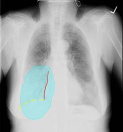
The opacity does not obscure the right heart border (red) but does obscure the right hemidiaphragm (yellow dotted line). |
||||||||||||