Case 1-Answers |
||||||||||
Imaging Pneumonia |
||||||||||
 |
||||
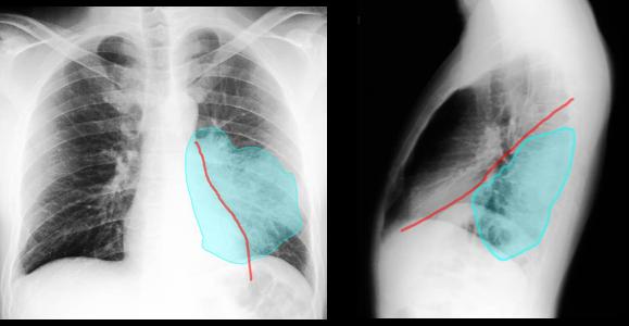
There is ill-defined opacity (white area) in the left posterior lower chest. This location suggests left lower lobe pneumonia. |
||||
 |
||||||
On the frontal view, the left heart border is still sharp, indicated in red (not obscured like it would be if the pneumonia were in the lingula). The area of consolidation is shown in blue. |
||||||
On the lateral view, the abnormality is all posterior to the major fissure, indicated in red. The area of consolidation is shown in blue. |
||||||