Case 10-Answers |
||||||||||||
Imaging Pneumonia |
||||||||||||
 |
||||||||||||
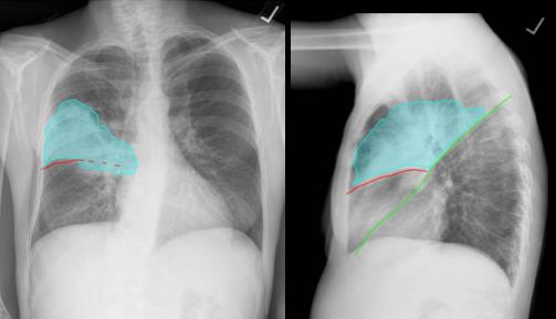
This consolidation is in the upper half of the right hemithorax on the frontal view and anterior in location on the lateral view, consistent with RUL pneumonia. |
||||||||||||
On the frontal view, the bottom edge of the area of opacity has a smooth margin, at least laterally, suggesting a fissure, in this case the minor fissure (red). The only lobe that is immediately superior to the minor fissure is the right upper lobe. |
||||||
 |
||||||
On the lateral view, the bottom edge of the area of opacity again abuts the minor fissure (red). The consolidation is large and also extends along the front edge of the major fissure (green). |
||||||