|
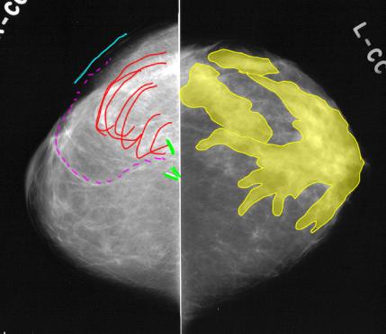
The left CC view is normal, with stroma that is denser than fat (shown in yellow) radiating out from the nipple region, and more stroma in the outer than the inner breast. On CC views, the lateral breast is always at the TOP of the image. On the right, there is no stroma in the region of the nipple, surgical clips (green), a skin marker indicating a scar (blue), and a swirling pattern in the outer breast (red), with a hint of some associated fat that seems to be separate from the rest of the breast (magenta dotted line). The lateral position of the swirling tissue does not fit with a TRAM reconstruction.
|
|
|