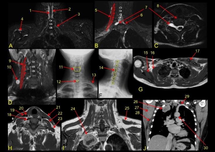
A-C are a patient s/p MVC with R UE sx |
||||||
 |
||||||
poster-answers |
||||||
 |
||||||||||||||||
Types of studies: B-MRI, T2w, FS, coronal C-MRI, T2w, FS, axial D-MRI, T2w, FS, coronal E-radiograph, AP F-radiograph, right oblique G-MRI, T1w, axial H-MRI, T1w, axial I-MRI, T1w, coronal J-CT, soft tissue, IV contrast, coronal FS=fat saturation T2w=T2-weighted T1w=T1-weighted |
||||||||||||||||
7-R C8 avulsion* 8-vertebral body 9-R anterior scalene 10- schwannoma 11- C5 vertebral body 12- C7 spinous process |
||||||||||||||||
1- spinal cord 2- CSF 3- L C6 nerve root 4-R supraspinatus 5-R middle scalene 6-L C8 nerve root |
||||||||||||||||
13-L first rib 14-R C4 neural foramen 15-R subscapularis 16- R serratus anterior 17-left clavicle fracture 18-R vertebral artery |
19-R internal jugular v 20-R com carotid a 21-L SCM 22-L anterior scalene 23-L middle scalene 24-Pancoast tumor
|
25-L trapezius 26-Pancoast tumor 27-R subscapularis 28-R serratus ant 29-L subclavian a 30-aortic arch
|
||||||||||||||
* with pseudomeningocele-outpouching of CSF space in the region of nerve root injury--the actual avulsed nerve is not visible, only the enlarged CSF space |
||||||||||||||||