|
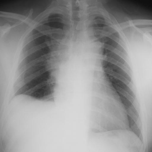
This case demonstrates 'lateralization of the peak' of the right diaphragm, as indicated by the arrow on the label marked 'abnormal contour'. This is a characteristic finding in subpulmonic pleural effusions, which typically do NOT show the expected blunting of the costophrenic angles we usually associate with pleural effusions. On the CT topogram (low resolution digital image done to plan where to image for a CT scan), the patient was supine and the fluid flowed out laterally. Most subpulmonic effusions are NOT loculated, meaning that the fluid collects inferior to the lung only temporarily, and can flow relatively freely into other positions as the patient moves around.
|
|