|
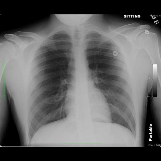
Pulmonary vascular redistribution is an early sign of CHF, and means that the diameter of vessels in the upper parts of the lung is increased. In a normal patient, as shown here, the upper lobe vessels should be very thin, much smaller in diameter than the lower lobe vessels. In addition, the outer 1/3 of the lung should have virtually no markings, as shown in this normal case. In CHF, either linear (if edema is involving the interstitium) or ill-defined alveolar opacities (if edema has flooded the alveoli) may be seen in the outer as well as more central parts of the lung.
|
|