Anatomy Yr-1 |
||||||||||||||
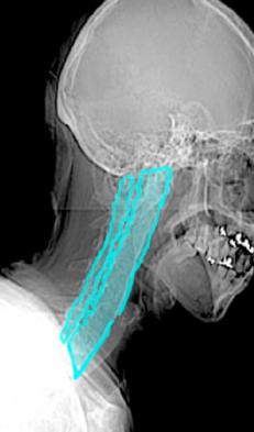
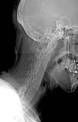
Image A looks like a poor quality lateral radiograph of the cervical spine, blurry and hard to interpret. It is actually a low quality digital image acquired at the start of a CT scan, called a 'topogram', 'scout', or 'scanogram'. It is used to help the technologist decide where to start and stop the scan. Image B shows the unusual appearance of the spine, with no disc spaces visible, indicated in aqua. The spine looks like one continuous column of bone. |
||||||
 |
||||||
 |
||||||
B
A
CASE 4