Tufts Anatomy Yr-1 |
||||||||||||||||||
This foot looks very washed-out, and the bones are hard to see overall. This suggests osteopenia or loss of bone mineralization. In the soft tissues are some faint linear calcifications that are in the walls of vessels. Small vessel damage is common in diabetes, and can show this appearance. Mouse over the labels to see the ulcer and the underlying bone. The concern is for an infection that has spread to the bone, called osteomyelitis. |
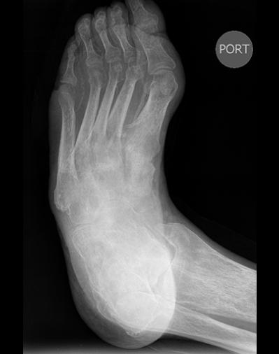 |
This is an AP projection, and the label 'PORT' suggests that was done as a portable study, suggesting the patient was too ill to come to the department for imaging. In general, whenever we do imaging of the limbs, we like to get at least TWO images at right angles to each other, so a lateral view would be indicated. |
||||||
red-digital arteries, |
||||||||