Tufts Anatomy Yr-1 |
||||||||||||
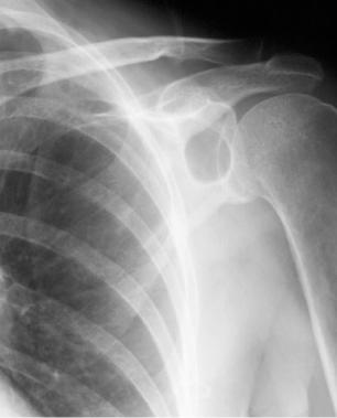
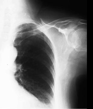
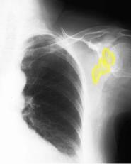
65 year old female with left shoulder pain |
||||||||||||
 |
||||||
 |
||||||
Image A is the closeup of our patient's left shoulder, and Image C shows in yellow a subtle area that is too dark (lucent), which is hard to see because of the angle that the x-ray beam is passing through the bony structures. Image B shows a left shoulder radiograph in our patient, lined up to better demonstrate the bones. The lucent area is much easier to see. Image D has outlines on structures of the shoulder region for you to identify. |
||||||

B
A
D
C
CASE 3 followup