PrISM Respiratory |
||||||||||||||
 |
||||
 |
||||
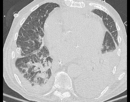
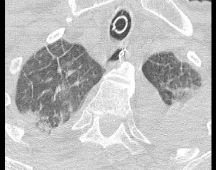
These two images are from the patient with cardiac history and severe shortness of breath. In the outermost lung there are many linear densities that are not visible on the normal CT (click the 'norma CTl' button to bring up the normal comparison images). These lines are due to fluid collecting in the interlobular septae of the lung, and indicate the presence of fluid in the intersitial space, which is between the alveoli and capillaries, leading to impaired diffusion of oxygen from alveoli to vessels and producing severe shortness of breath.
The thickened interlobular septae show the basic structure of the lung, which is not normally visible. The lung is organized into lobules that are polygonal in shape and contain a pulmonary arterial branch in the center (which appears on the scans as a dot). There are also hazy areas of ground glass opacity filling the alveoli and smaller interstitial structures of some of the lobules, indicating more severe edema.