PrISM Respiratory |
||||||||||||||
 |
||||
 |
||||
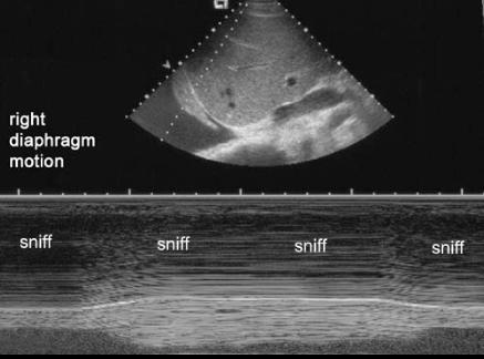
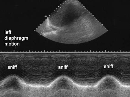
This is an example of a real-time way to visualize the actual motion of the diaphragm with ultrasound in a different patient. It is called a 'sniff test', and is done by placing the ultrasound probe in such a location as to visualize first one and then the other diaphragm (shown in the labeled images in yellow-these images are oriented in the sagittal plane). Then a region of the image is analyzed (indicated in blue) and displayed in a read-out at the bottom that shows how much the diaphragm moves when you ask the patient to sniff. In this case, the left diaphragm moved normally with sniffing, but the right diaphragm did not move at all. Sometimes, in a completely paralyzed diaphragm, when the patient sniffs, the abnormal side may actually go UP instead of DOWN (paradoxical motion--not seen in this case).