PrISM Respiratory |
||||||||||||||
 |
||||
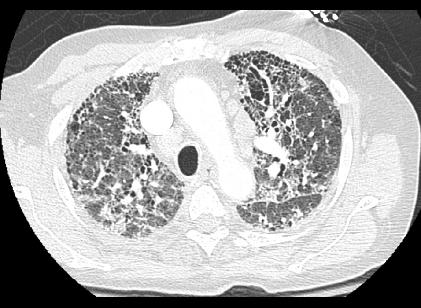
This axial CT at a level similar to the prior case shows a very different appearance of the lungs, with many small holes lined up in rows around the edges of the lung and outlining many of the vascular structures. There are also areas of hazy opacity (ground-glass) and extra lines. This is a typical appearance of diffuse lung fibrosis and the PFTs would be expected to show low lung volumes and a restrictive pattern.
A coronal reconstruction of the CT data can be particularly helpful in showing where the disease is worst. In this case, the posterior and basal lungs are slightly more abnormal than other regions, but no part of the lung is spared.