PrISM Respiratory |
||||||||||||||
 |
||
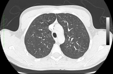
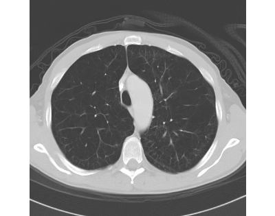
The image on the left is from this patient, and the image on the right is a normal comparison study.
Normal patient
This patient's lungs are too lucent on CT as well, with many black holes of varying size, compared to the normal faintly grey appearance of healthy lung. There are not as many branching vessels as normal, and the vessels that are present appear stretched and distorted by the surrounding lucent areas, or bullae. The trachea is narrow from side to side and appears compressed (sabre sheath trachea). This is the typical imaging appearance of emphysema, and an obstructive pattern would be expected on pulmonary function testing (PFT) in this patient.