PrISM Cardiovascular |
||||||||||||||
 |
 |
|||||||||||||
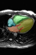
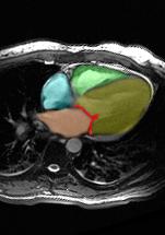
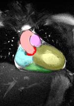
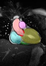
These are MR images of the heart in the axial (Images A and B) and coronal (Images C and D) planes. Images A and C are in ventricular diastole and Images B and D are in ventricular systole. The right ventricle is in green, the left ventricle in yellow, the right atrium in aqua and the left ventricle in orange. The mitral valve on Image A is in red. The aortic valve in Image B is in red. The aorta is in pink and the main pulmonary artery in purple on Images C and D. There is an abnormally high ejection fraction in this case, with near complete obliteration of the left ventricular chamber at systole. This is due to left ventricular hyptertophy or thickening of the left ventricular wall, which can be idiopathic or can be related to hypertension or aortic valve stenosis. |
||||||||||||||
image B |
||||||||||||||
image A |
||||||||||||||
 |
 |
|||||||||||||
image D |
||||
image C |
||||