PrISM Cardiovascular |
||||||||||||||||||||||||||||||||||
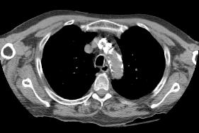
A |
||||||||||||||||||||||||||||||||||
 |
||||||||||||||||||||||||||||||||||
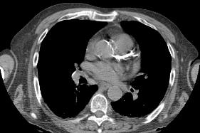
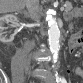
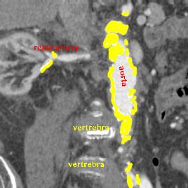
The previous images were axial CT scans in soft tissue windows. It is normal to see the aorta on a CT scan, unlike a radiograph, because CT is much more sensitive to slight differences in tissue density. However the aorta on this study is NOT normal, but is very densely calcified. This is a sign of severe atherosclerosis, sometimes called a 'coral reef' aorta, because if the surgeon attempts to correct the blockage by sewing in a graft, it is like trying to sew into a rock. Selected images are shown on the right, with labels to indicate the calcifications. Images A and B are done without IV contrast. Image C is a coronal reformatted image with IV contrast. |
||||||||||||||||||||||||||||||||||
 |
||||||||||||||||||||||||||||||||||
B |
||||||||||||||||||||||||||||||||||
 |
||||||||||||||||||||||||||||||||||
C |
||||||||||||||||||||||||||||||||||
NOAA photo library
yellow=abnormal calcium deposits in walls of arteries
ao=aorta, b=brachiocephalic artery, c=left common carotid artery
LAD=left anterior descending coronary artery