PA Anatomy: CN/spine: Case 1 |
||||||
 |
||||
 |
||||
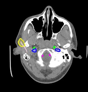
This is a normal axial CT image, with a some structures labeled. The pink indicates the normal position of CN VII, entering the parotid gland (shown in yellow), after exiting the skull through the stylomastoid foramen. Carotids are in green, internal jugular veins in dark blue, and vertebral arteries in purple. |
|||||||||||||||
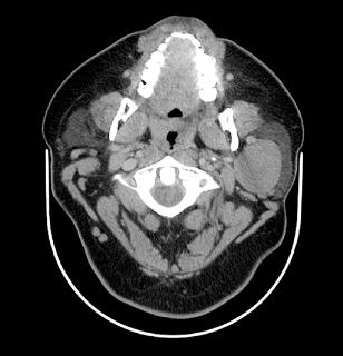
This patient has a tumor of the left parotid gland, which is compressing or damaging CN VII (facial). This would explain facial weakness. The imaging is a axial CT with soft tissue windows and very faint IV contrast (not enough given for her weight, which was over 400 pounds). |
||