PA Anatomy: CN/spine: Case 1 |
||||||
 |
||||
 |
||||
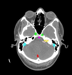
This is a normal axial CT image, with a some structures labeled. The pink indicates the normal position of CN VIII, entering the internal acoustic meatus (shown in yellow), accompanied by the CN VII. Carotids are in green, sigmoid sinuses in blue, basilar artery in purple, and occipital sinus in red. |
||||||||||
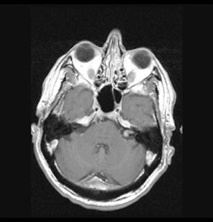
This patient has a tumor of CN VIII, the vestibulocochlear nerve. This would explain hearing loss. The imaging is a axial MR with T1 weighting and using gadolinium contrast. |
||