PA Anatomy: CN/spine: Case 1 |
||||||
 |
||||
 |
||||
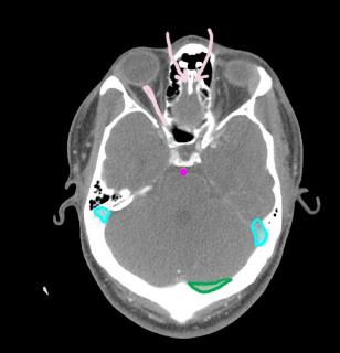
This is a normal axial CT image, with a few structures labeled. The pink arrows point to the normal location of the olfactory nerve bulb. The pink line is CN II, the optic nerve. Venous sinuses are labeled in blue (sigmoid) and green (transverse). The basilar artery is labeled in purple |
||||||||
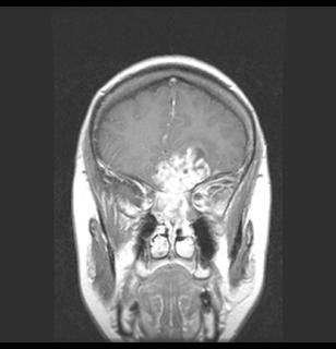
red-normal cerebral vessels, showing that contrast was used; blue-tumor of CN I; yellow-normal level of cribriform plate |
||||||||
This patient has a tumor of CN 1, the olfactory nerve. This would explain her loss of her sense of smell. The imaging is a coronal MR with T1 weighting and using gadolinium contrast. |
||