PA Anatomy: Brain: Case 2 |
||||||
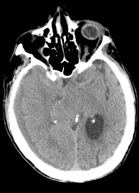
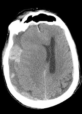
Streaks on the lower images are due to dental work (metal artifact). The black streaks on some of the upper images are due to motion. The patient was not holding still during the scan. This is a CT in the axial plane without IV contrast |
||||||
 |
 |
|||||
Because acute hemorrhage usually appears whiter than normal vessels or brain tissue, we often do CT scanning initially after trauma WITHOUT IV contrast. In this way, we can increase our sensitivity for detecting small amounts of blood (white areas). Do you see anything you think is fresh blood on these images? |
||